
MICROSCOPIC URINALYSIS

Methodology
A sample of well-mixed urine (usually
10-15 ml) is centrifuged in a test tube at relatively low speed (about 2-3,000
rpm) for 5-10 minutes until a moderately cohesive button is produced at the
bottom of the tube. The supernatant is decanted and a volume of 0.2 to 0.5 ml
is left inside the tube. The sediment is resuspended in the remaining
supernatant by flicking the bottom of the tube several times. A drop of
resuspended sediment is poured onto a glass slide and coverslipped.
Examination
The sediment is first examined under low power to identify most crystals,
casts, squamous cells, and other large objects. The numbers of casts seen are
usually reported as number of each type found per low power field (LPF).
Example: 5-10 hyaline casts/L casts/LPF. Since the number of elements found in
each field may vary considerably from one field to another, several fields are
averaged. Next, examination is carried out at high power to identify crystals,
cells, and bacteria. The various types of cells are usually described as the
number of each type found per average high power field (HPF). Example: 1-5
WBC/HPF.
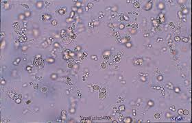
Red Blood Cells
Hematuria is the presence of abnormal numbers of red cells in urine due to: glomerular damage, tumors which
erode the urinary tract anywhere along its length, kidney trauma, urinary tract
stones, renal infarcts, acute tubular necrosis, upper and lower uri urinary
tract infections, nephrotoxins, and physical stress. Red cells may also
contaminate the urine from the vagina in
menstruating women or from trauma produced by bladder catherization.
Theoretically, no red cells should be found, but some find their way into the
urine even in very healthy individuals.
However, if one or more red cells can be found in every high power field, and
if contamination can be ruled out, the specimen is probably abnormal.

RBC's may appear normally shaped, swollen by
dilute urine (in fact, only cell ghosts
and free hemoglobin may remain), or crenated by concentrated urine . Both
swollen, partly hemolyzed RBC's and crenated RBC's are sometimes difficult to
distinguish from WBC's in the urine . In addition, red cell ghosts may simulate
yeast. The presence of dysmorphic RBC's in
urine suggests a glomerular disease such as a glomerulonephritis.
Dysmorphic RBC's have odd shapes as a consequence of being distorted via
passage through the abnormal glomerular structure.
White Blood Cells
Pyuria refers to the presence of abnormal numbers of leukocytes that may
appear with infection in either the upper or lower urinary tract or with acute
glomerulonephritis. Usually, the WBC's are granulocytes. White cells from the
vagina, especially in the presence of vaginal and cervical infections, or the
external urethral meatus in men and women may contaminate the urine .

If two or more leukocytes per each high power field appear in
non-contaminated urine , the specimen is probably abnormal. Leukocytes have
lobed nuclei and granular cytoplasm.
Epithelial Cells
Renal tubular epithelial cells, usually larger than granulocytes, contain a
large round or oval nucleus and normally slough into the urine in small numbers. However, with nephrotic
syndrome and in conditions leading to tubular degeneration, the number sloughed
is increased.


When lipiduria occurs, these cells contain endogenous fats. When filled with
numerous fat droplets, such cells are called oval fat bodies. Oval fat bodies
exhibit a "Maltese cross" configuration by polarized light microscopy.
Transitional epithelial cells from the renal pelvis, ureter, or bladder have
more regular cell borders, larger nuclei, and smaller overall size than
squamous epithelium. Renal tubular epithelial cells are smaller and rounder
than transitional epithelium, and their nucleus occupies more of the total cell
volume.

Squamous epithelial cells from the skin surface or from the outer urethra
can appear in urine .

Their significance is that they represent
possible contamination of the specimen with skin flora.
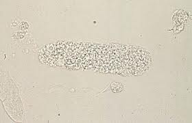
Casts
Urinary casts are formed only in the distal convoluted tubule (DCT) or the
collecting duct (distal nephron). The proximal convoluted tubule (PCT) and loop
of Henle are not locations for cast formation. Hyaline casts are composed
primarily of a mucoprotein (Tamm-Horsfall protein) secreted by tubule cells.
The Tamm-Horsfall protein secretion (green dots) is illustrated in the diagram
below, forming a hyaline cast in the collecting duct:
Even with glomerular injury causing increased glomerular permeability to
plasma proteins with resulting proteinuria, most matrix or "glue"
that cements urinary casts together is Tamm-Horsfall mucoprotein, although
albumin and some globulins are also incorporated. An example of glomerular
inflammation with leakage of RBC's to produce a red blood cell cast is shown in
the diagram below:
The factors which favor protein cast formation are low flow rate, high salt
concentration, and low pH, all of which favor protein denaturation and
precipitation, particularly that of the Tamm-Horsfall protein. Protein casts with
long, thin tails formed at the junction of Henle's loop and the distal
convoluted tubule are called cylindroids. Hyaline casts can be seen even in
healthy patients.
Red blood cells may stick together and form red blood cell casts. Such casts
are indicative of glomerulonephritis, with leakage of RBC's from glomeruli, or
severe tubular damage.

White blood cell casts are most typical for acute pyelonephritis, but they
may also be present with glomerulonephritis. Their presence indicates
inflammation of the kidney, because such casts will not form except in the
kidney.


When cellular casts remain in the nephron for some time before they are
flushed into the bladder urine , the cells may degenerate to become a coarsely
granular cast, later a finely granular cast, and ultimately, a waxy cast.
Granular and waxy casts are be believed to derive from renal tubular cell
casts. Broad casts are believed to emanate from damaged and dilated tubules and
are therefore seen in end-stage chronic renal disease.
 granular cast
 Waxy Cast
The so-called telescoped urinary sediment is one in which red cells, white
cells, oval fat bodies, and all types of casts are found in more or less equal
profusion. The conditions which may lead to telescoped sediment are: 1) lupus
nephritis 2) malignant hypertension 3) diabetic glomerulosclerosis, and 4)
rapidly progressive glomerulonephritis.
In end-stage kidney disease of any cause, the
urinary sediment often becomes very scant because few remaining nephrons
produce dilute urine
Bacteria

Bacteria are common in urine
specimens because of the abundant normal microbial flora of the vagina or
external urethral meatus and because of their ability to rapidly multiply
in urine standing at room temperature.
Therefore, microbial organisms found in all but the most scrupulously collected
urines should be interpreted in view of clinical symptoms.
Diagnosis of bacteriuria in a case of suspected urinary tract infection
requires culture. A colony count may also be done to see if significant numbers
of bacteria are present. Generally, more than 100,000/ml of one organism
reflects significant bacteriuria. Multiple organisms reflect contamination.
However, the presence of any organism in catheterized or suprapubic tap
specimens should be considered significant.
Yeast
Yeast cells may be contaminants or represent a true yeast infection. They
are often difficult to distinguish from red cells and amorphous crystals but
are distinguished by their tendency to bud. Most often they are Candida, which
may colonize bladder, urethra, or vagina.
Crystals
Common crystals seen even in healthy patients include calcium oxalate,
triple phosphate crystals and amorphous phosphates.


Very uncommon crystals include: cystine
crystals in of neonates with congenital
urine cystinuria or severe liver disease, tyrosine crystals with
congenital tyrosinosis or marked liver impairment, or leucine crystals in
patients with severe liver disease or with maple syrup urine disease.
Miscellaneous
General "crud" or unidentifiable objects may find their way into a
specimen, particularly those that patients bring from home.
Spermatozoa can sometimes be seen. Rarely, pinworm ova may contaminate the
urine In Egypt, ova from bladder
infestations with schistosomiasis may be seen.